What Ten Days of Bed Rest Did to Aging Mitochondria
Prefer to listen? Hit play for a conversational, audio‑style summary of this article’s key points.
Ten days of bed rest left older adults' mitochondrial energy engines intact. In a detailed study of ten healthy older men, published in the Journal of Physiology, mitochondrial respiration was fully preserved after ten days of strict bed rest, under both moderate and maximal demand. The protein machinery of energy production was unchanged too. The intuitive assumption that disuse weakens the muscle by breaking its energy-producing mitochondria did not hold.
The remaining mitochondria actually became slightly more efficient. Because there were fewer mitochondria after bed rest, the researchers measured how well each individual one performed and found intrinsic efficiency had risen. The muscle appeared to compensate for a smaller mitochondrial population by getting more out of each remaining unit, a sign of the aging muscle adapting rather than simply failing.
What did decline was the number of mitochondria. Electron microscopy showed reduced mitochondrial volume density, meaning fewer mitochondria occupied the muscle. Gene-expression data pointed to why: a coordinated downregulation of the genes that build mitochondria alongside activation of the recycling pathway that clears worn-out ones, suggesting the muscle was removing mitochondria faster than it replaced them. This recycling link is probable rather than proven, since the key recycling proteins themselves did not change.
Oxidative stress rose, but not because the engines got dirtier. The mitochondria emitted more reactive oxygen species after bed rest, but only under conditions when they were not being driven to make energy. When signaled to produce ATP, they controlled their exhaust normally. This is a crucial distinction: the engines were not malfunctioning or leaking more waste for their condition.
The real problem was a weakened defense against that exhaust. The rise in oxidative stress traced to a dysregulation of the muscle's antioxidant defenses, the enzyme systems that neutralize reactive oxygen species, including the downregulation of GPX1, a frontline enzyme. The oxidative stress rose because the cleanup system was turned down, not because the mitochondria produced more waste. The authors emphasize that this redox imbalance occurred upstream of, and independent of, any failure in energy production.
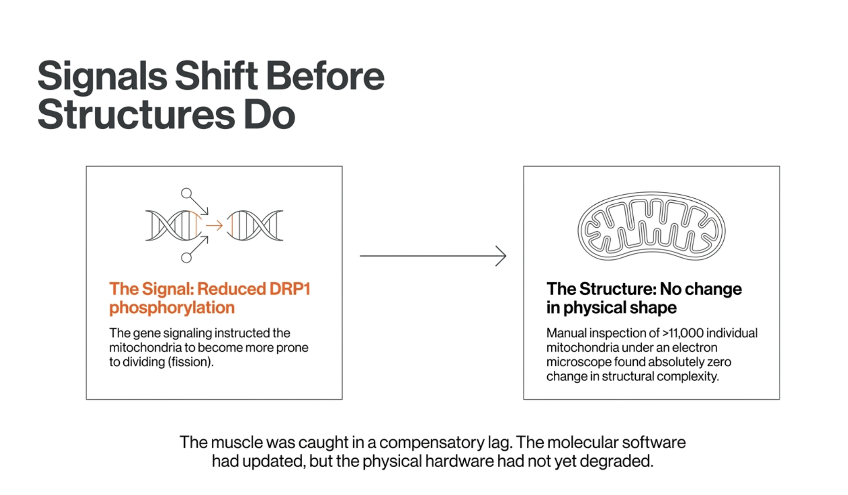
The layers of mitochondrial biology did not move together. The study's recurring theme is that the signals and genes shifted while the functional and structural reality lagged. The signaling that governs mitochondrial division changed (reduced DRP1 phosphorylation, which should favor fission), yet manual inspection of more than eleven thousand individual mitochondria found no change in their actual shape. The muscle was caught in an early, still-compensating phase of a response that had not fully played out.
If the engines held, why do older adults get weaker after bed rest? Likely the cardiovascular system. The authors reason that since muscle mitochondria can still use oxygen to make energy, the loss of whole-body exercise capacity after bed rest probably stems from earlier in the oxygen-delivery chain, the heart, blood volume, and the microvascular and endothelial networks that decondition quickly during inactivity. This is an inference from finding the mitochondria intact, not a direct measurement in this study.
This reframing is, in a sense, encouraging. Failing energy engines would be a deep and hard-to-reverse problem. A redox imbalance, a thinned mitochondrial population, and cardiovascular deconditioning are all things the body is well equipped to reverse, and the study captured an early, largely reversible phase. All three respond to the same intervention: physical activity.
It points to movement, not antioxidant supplements. It is tempting to read "oxidative stress rose" as a reason to take antioxidants, but the research on that is discouraging, high-dose antioxidant supplements have generally failed to help and can even blunt the beneficial adaptations exercise produces. The body's own antioxidant systems, mitochondrial replacement, and cardiovascular conditioning are best supported by using the muscle. The practical lesson is to avoid unnecessary bed rest and to get older people moving early and safely during and after illness.
The findings are real but bounded, and the deepest lesson is about invisibility. The study was small (ten men), all male, acute (ten days, a single post-bed-rest snapshot), and several key conclusions rest on gene expression rather than measured function. But its core finding is well supported, and it is a vivid reminder that the molecular changes of aging and disuse happen silently, detectable only by measuring them, long before they surface as weakness. Knowing where your body actually stands, rather than guessing from how you feel, is the point of measuring your biology.
Introduction: What Ten Days in Bed Does to an Aging Body
It is one of the quiet dangers of growing older, and it rarely gets the attention it deserves. An older adult catches a bad case of the flu, breaks a bone, or spends a week in a hospital bed recovering from a procedure, and emerges from that short stretch of enforced stillness noticeably weaker. Sometimes the strength comes back. Sometimes it does not, and that brief period in bed becomes the start of a longer decline toward frailty and lost independence. Clinicians have long known that short-term inactivity is harder on older bodies than younger ones, and that the muscle loss it causes can be strikingly fast.
The standard assumption about why has centered on the mitochondria, the tiny power plants inside muscle cells. Disuse, the thinking goes, damages these engines, leaving the muscle less able to produce energy, and that energy failure is what weakens it. It is an intuitive story, and mitochondrial decline is genuinely a feature of both aging and inactivity. But a new study set out to look directly at what ten days of bed rest actually does to the mitochondria of older adults, in unusual molecular detail, and the picture it found is not the one the standard story predicts.
The study, published in the Journal of Physiology, put ten healthy older men through ten days of strict bed rest, a deliberate stand-in for the kind of short hospitalization or illness that so often triggers decline. Before and after, the researchers took muscle biopsies and examined them from every angle they could: how well the mitochondria produced energy, how much oxidative stress they generated, how many of them there were, what they looked like under an electron microscope, and which genes had switched on or off. Few studies of disuse have looked this comprehensively at the same tissue.
What they found reframes the problem. The mitochondrial engines did not break. Energy-producing capacity was fully preserved, and by one measure each remaining mitochondrion was actually working slightly better. The damage ten days of stillness did to these older muscles was real, but it was somewhere else entirely: in a rising tide of oxidative stress that the muscle's antioxidant defenses were no longer keeping in check, and in a quiet thinning of the mitochondrial population. The engines ran fine. What faltered was the system meant to neutralize their exhaust.
That distinction is not academic. It changes what you would do to protect an aging body through a period of forced rest, and it points, as we will see, to a culprit for post-illness weakness that may lie outside the muscle altogether. What follows is what ten days in bed did and did not change, why oxidative stress rose even as the mitochondria kept working, and what it suggests about protecting muscle, and independence, across the vulnerable stretches of later life.
Mitochondria Do More Than Make Energy
To understand what the study found, it helps to let go of the idea that mitochondria are simply the cell's power plants. That is their most famous job, but it is only one of several, and the others turn out to be central to this story.
The power-plant role is real and it is enormous. Mitochondria take in oxygen and fuel and run them through a process called oxidative phosphorylation to produce ATP, the molecule that powers essentially everything a muscle cell does, above all, contraction. A muscle working hard burns through staggering quantities of ATP, and the mitochondria are what regenerate it. When people talk about mitochondrial decline weakening a muscle, this is the function they have in mind: fewer or feebler engines, less energy, a weaker muscle.
But mitochondria are also, unavoidably, a source of exhaust. Producing energy with oxygen generates reactive oxygen species, or ROS, chemically reactive molecular fragments that are an inevitable byproduct of burning fuel. In moderation, ROS are not merely waste; at low levels they serve as useful signals that help the cell adapt and respond. In excess, though, they damage proteins, membranes, and DNA, a state called oxidative stress. Healthy cells manage this with a dedicated antioxidant defense system, a set of enzymes whose job is to neutralize ROS and keep it in check. The exhaust is constant; staying healthy depends on continuously cleaning it up.
Mitochondria also maintain themselves, and the cell maintains them, through quality control. Worn-out or damaged mitochondria are identified and recycled through a process called mitophagy, a kind of cellular waste disposal that clears out failing units so they can be replaced. This is a different housekeeping system from the antioxidant defenses, and the distinction matters for this study: one system neutralizes the chemical exhaust the mitochondria produce, the other removes the spent mitochondria themselves. A healthy muscle needs both running well.
These functions matter especially in older adults, and they matter most precisely when the body is forced to rest. Aging already erodes mitochondrial quality control and antioxidant capacity, leaving less margin. Inactivity adds a second stress on top of that, and older bodies have a harder time absorbing it. A younger person can lose ground during a week in bed and regain it quickly; an older person often loses more and recovers less completely. So when researchers wanted to know what short-term disuse does to the aging muscle, the question was not only whether the energy engines falter, but what happens across all of these jobs at once, the energy production, the exhaust management, and the recycling. That is what made this study's all-angles approach worth doing.
The Study
The design was built to answer a clinically grounded question, and the choice of timeframe was deliberate. Many disuse studies use shorter stints of a few days or much longer immobilizations of a month or more. These researchers chose ten days because it mirrors something that happens constantly in the real world: the length of a typical hospital stay, a recovery from illness, or a stretch of bed-bound convalescence that an older adult might go through several times across their later years. Ten days is not an extreme scenario. It is an ordinary one, which is part of what makes the findings matter.
Ten healthy older men, average age about 68, spent ten days in strict horizontal bed rest in a hospital setting. Strict is the operative word: they remained lying down throughout, with no muscle-contracting activity permitted, so the study could isolate the effect of disuse itself rather than of reduced exercise layered onto ordinary movement. Their diet was controlled and calibrated to their energy needs, so weight loss or overfeeding would not confound the results. This is a clean, if demanding, model of what pure inactivity does to an aging body.
The real strength of the study is what the researchers did with the muscle. They took biopsies from the thigh before the bed rest and again after the tenth day, and then examined the same tissue through an unusually complete set of methods, each a different window onto the mitochondria. High-resolution respirometry measured how well the mitochondria actually produced energy, under both moderate and maximal demand. A separate assay measured how much reactive oxygen species the mitochondria emitted, the exhaust. Protein analysis quantified the key machinery of energy production, of mitochondrial fusion and fission, and of recycling. Electron microscopy let them see the mitochondria directly, counting how much of the muscle's volume they occupied and inspecting their shape and structure. And RNA sequencing read out which of thousands of genes had been switched up or down, revealing the muscle's programmed response at its deepest level.
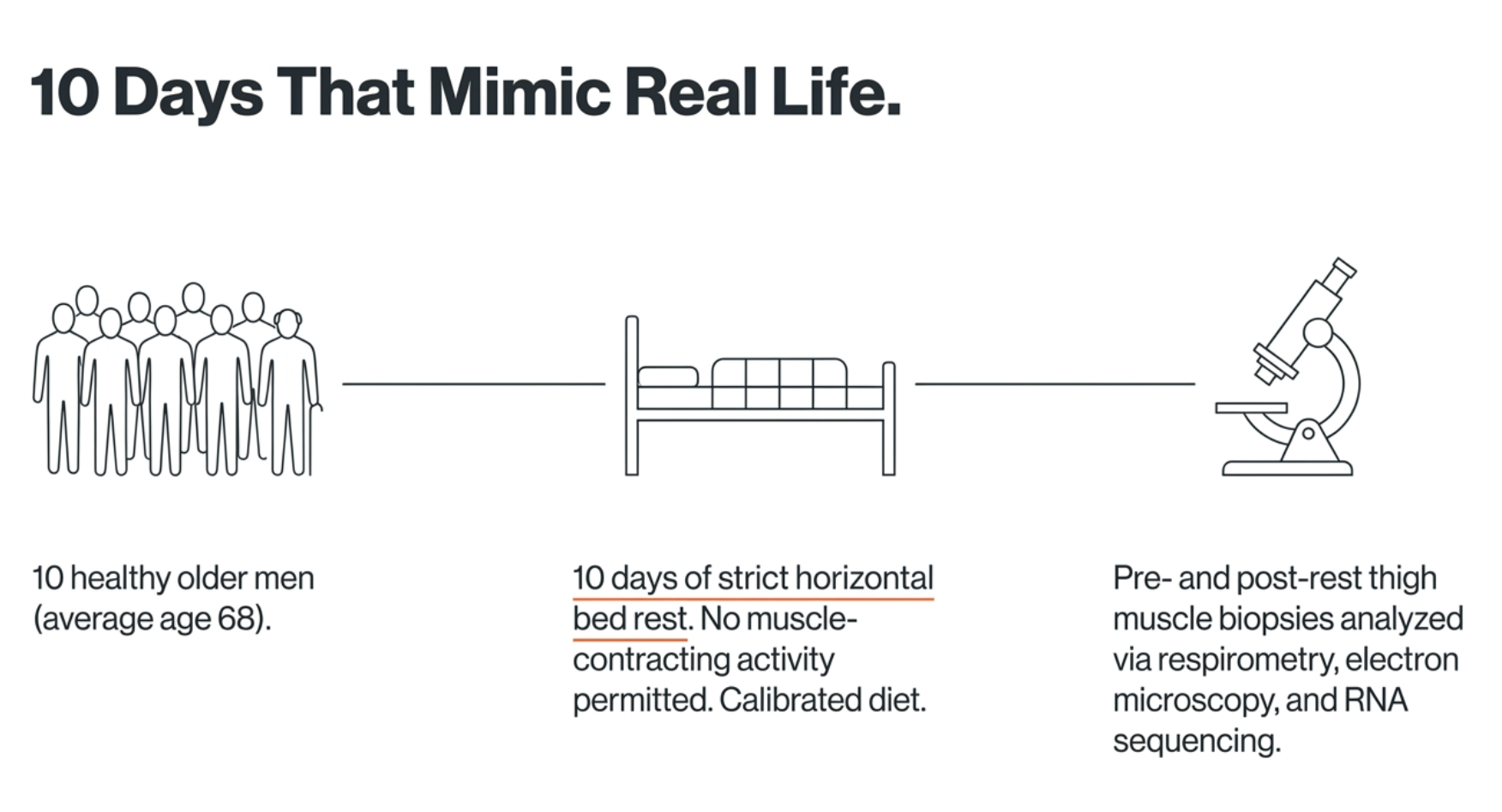
Few studies of disuse bring all of these tools to bear on the same tissue at once, and that is what gives this one its value. Any single measure can mislead. Energy output might look fine while the underlying machinery is quietly degrading, or the gene-expression program might be shifting in ways that haven't yet shown up in function. By triangulating across function, structure, protein, and gene expression simultaneously, the researchers could see not just whether something changed, but where in the chain of cause and effect the change actually sat. That turned out to be the whole point, because the different layers did not all tell the same story.
The First Surprise: The Engine Didn't Break
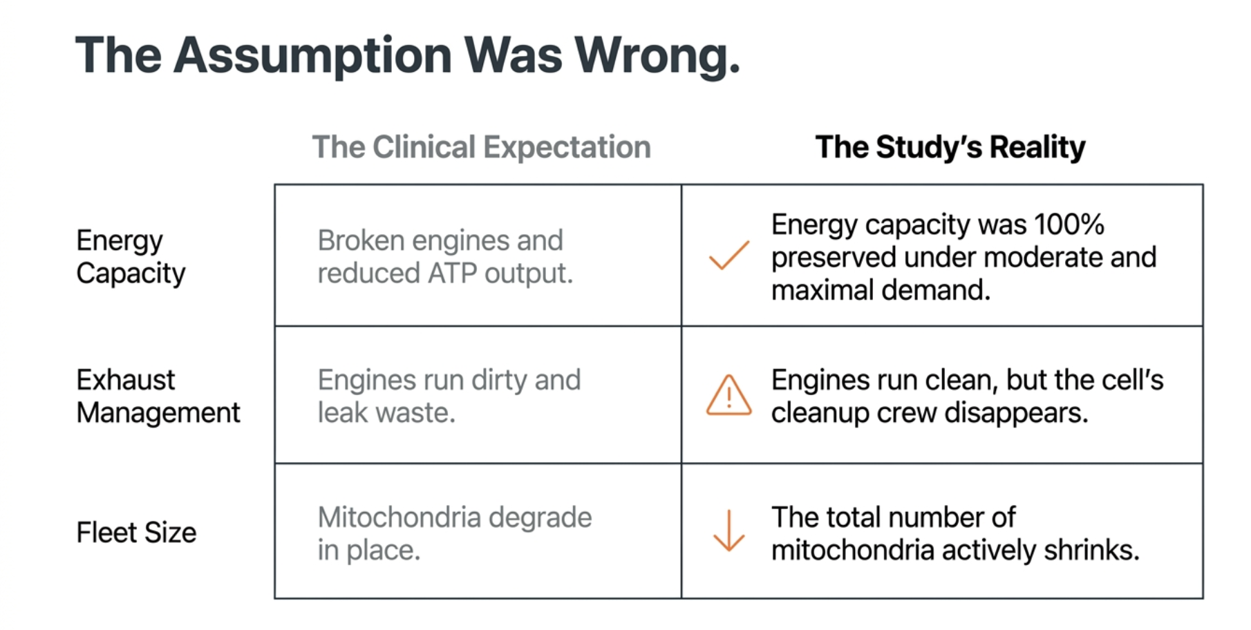
The expectation going in was straightforward. Ten days of disuse in older muscle should degrade mitochondrial function, leaving the engines weaker and the muscle less able to produce energy. That is the story disuse is supposed to tell. It is not the story the respirometry told.
When the researchers measured how well the mitochondria produced energy, under both moderate and maximal demand, they found no decline. Respiratory capacity was fully preserved after ten days in bed. The mitochondria were making energy just as well at the end of the bed rest as they had been at the start. By the single most direct measure of whether the engines still worked, the disuse had done nothing to them.
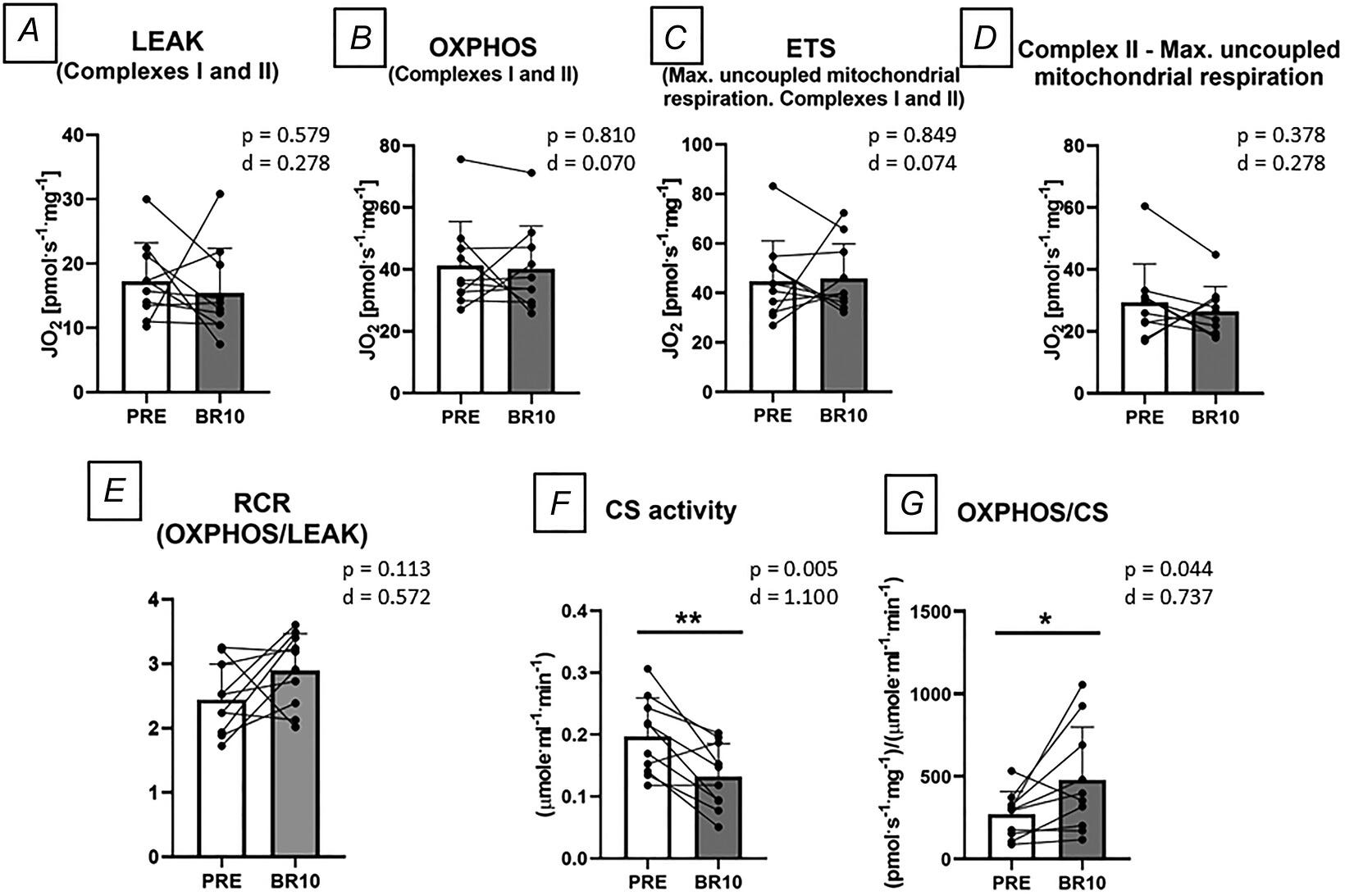
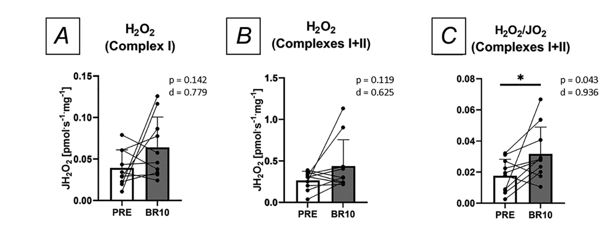
Figure 1: The engine held; the exhaust rose. Mitochondrial respiration was unchanged after 10 days of bed rest, under both moderate and maximal demand. Reactive oxygen species emission increased, specifically when the mitochondria were not being driven to make energy, pointing to weakened antioxidant clearance rather than dirtier engines.
Then came a result that pushed past merely preserved into mildly counterintuitive. When the researchers accounted for the fact that there were now fewer mitochondria in the muscle, more on that shortly, and asked how well each individual mitochondrion was performing, the answer was that intrinsic efficiency had actually gone up. Per unit of mitochondria, the engines were running slightly better than before. The muscle appeared to be compensating for having fewer mitochondria by getting more out of each one that remained.
The protein evidence backed this up. The machinery of energy production, the respiratory complexes and the larger assemblies called supercomplexes that carry out oxidative phosphorylation, showed no significant loss. The engines were not just performing well in the functional test; the physical components that make up those engines were still present and intact. Function and hardware agreed: the energy-production apparatus had weathered ten days of stillness without meaningful damage.
This is a genuine departure from what some earlier work predicted. At least one prior bed-rest study in older adults had found that disuse reduced both the amount of energy-production machinery and the mitochondria's functional output. Here, neither fell. The researchers are careful about the discrepancy, noting that detecting modest protein changes in human muscle is technically difficult and that biological variability between studies is large. But the broader point they draw is the important one: the different layers of mitochondrial biology do not always move together. Energy output, the amount of machinery, and the number of mitochondria can each change, or fail to change, on their own schedule. In this case, the function held even as other things shifted underneath it.
So if the engines were intact, even modestly improved per unit, then whatever was going wrong in these older muscles after ten days in bed, and something was going wrong, was not a failure of energy production. It was happening elsewhere. Two things, in fact, had changed, and finding them required looking past the respiration data to the exhaust and the fleet.
What Did Change: A Thinning Fleet and Rising Exhaust
If the engines held, two other things did not, and together they are where the real damage of ten days in bed showed up.
The first was the number of mitochondria. Using electron microscopy to look directly at the muscle tissue, the researchers measured mitochondrial volume density, the fraction of the muscle's interior actually occupied by mitochondria, and found it had fallen after the bed rest. There were simply fewer mitochondria in the muscle than before. The fleet had thinned, even though each remaining vessel was running well. This is why the earlier efficiency finding mattered: the muscle was getting slightly more out of each mitochondrion, which looks like a compensation for having lost some of them.
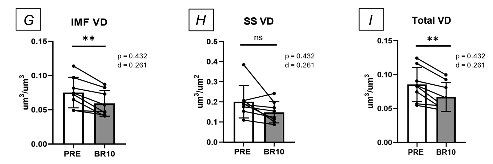
Figure 2: Fewer mitochondria after bed rest. Electron microscopy showed a reduction in mitochondrial volume density, the fraction of the muscle occupied by mitochondria, indicating a smaller mitochondrial population. The remaining mitochondria, however, kept the muscle's energy production intact.
The gene-expression data pointed to why the fleet shrank. Among the more than three thousand genes whose activity changed over the ten days, the researchers saw a coordinated downregulation of genes involved in building and running the mitochondria, the machinery of oxidative phosphorylation and the structural components of the organelle itself. At the same time, the recycling pathway showed signs of activation: several of the cargo-tagging genes that mark worn-out mitochondria for disposal were switched up. The combination, less building alongside more flagging-for-removal, fits a picture in which the muscle was clearing out mitochondria faster than it was replacing them, which would reduce the total count. The researchers are careful here, because the key recycling proteins themselves did not change at the protein level even as the genes did, so they present altered recycling as a likely contributor to the thinning fleet rather than a proven one.
There was one more change worth noting, because it reinforces how unevenly the different layers of mitochondrial biology responded. Mitochondria constantly divide and fuse, splitting into smaller units (fission) or merging into larger networks (fusion), and the balance between these shapes the health of the population. After bed rest, the researchers found a change in the signaling that governs this balance: reduced phosphorylation of a protein called DRP1 at a specific site, a shift that would be expected to make the mitochondria more prone to fission, to dividing. But when they went looking for the physical consequence, manually inspecting the shape of more than eleven thousand individual mitochondria under the electron microscope, they found none. The mitochondria had not actually changed shape: there was no measurable difference in how elongated, branched, or structurally complex they were. The instruction to divide more readily had shifted, but the visible structure had not yet followed. It is one more instance of the study's recurring theme, that after ten days in bed, the signals and the genes were moving while the functional and structural reality lagged behind, a muscle caught in the early, still-compensating phase of a response that had not yet fully played out.
The second major change was the one the authors treat as most important: oxidative stress rose. After ten days in bed, the mitochondria emitted more reactive oxygen species, more exhaust. But the details of when and why are what make this finding meaningful, and they point away from the engines.
The rise in ROS appeared specifically under what are called non-phosphorylating conditions, when the mitochondria were not actively being driven to make ATP. Critically, when the researchers supplied the signal that tells mitochondria to produce energy, the excess ROS was suppressed normally, exactly as it should be. This tells you something precise: the machinery that couples energy demand to exhaust control was still working. The engines were not leaking more exhaust because they were damaged or running dirty. Something else was letting the exhaust accumulate.
That something else was the cleanup. The gene-expression data revealed a dysregulation of the muscle's antioxidant defenses, the enzyme systems whose job is to neutralize ROS. Key antioxidant and oxidoreductase genes were altered, including the downregulation of GPX1, one of the frontline enzymes for clearing hydrogen peroxide, a major form of ROS. In other words, the mitochondria were producing roughly the normal amount of exhaust for their condition, but the system responsible for mopping it up had been turned down. The oxidative stress rose not because the engines got dirtier, but because the cleanup crew was understaffed.
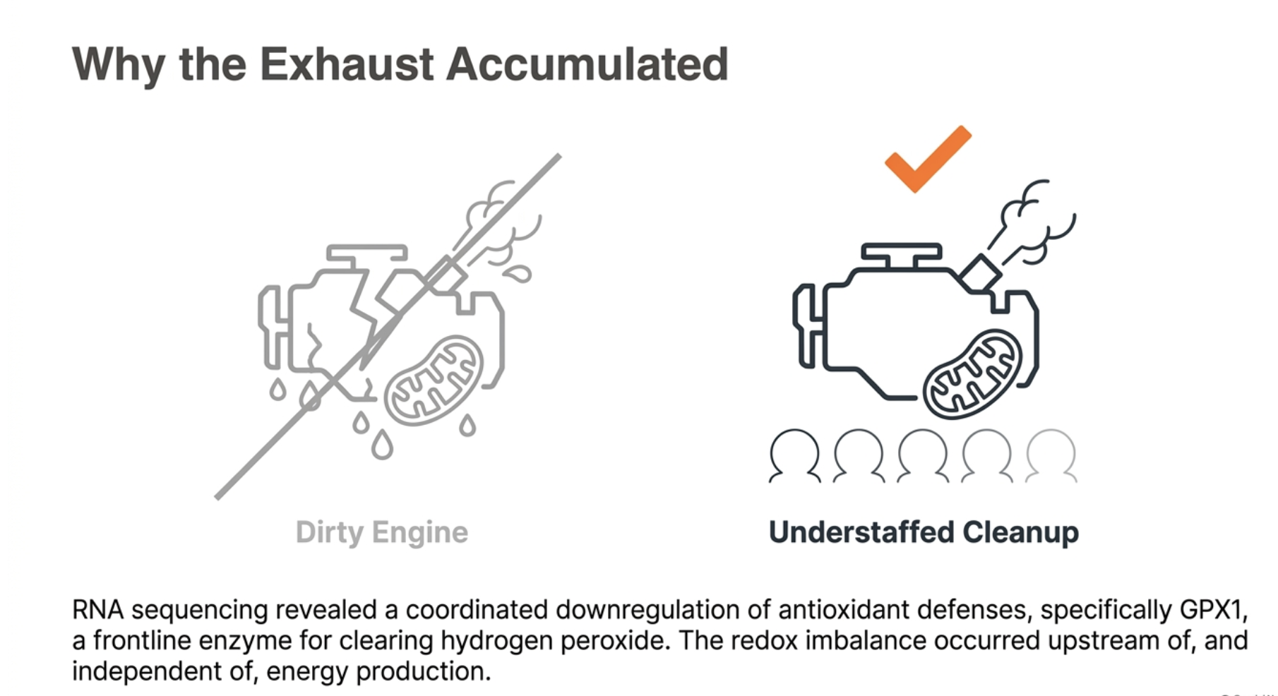
This is the finding that reframes the whole problem, and the authors state it plainly: the rise in oxidative stress occurred upstream of, and independent of, any impairment in energy production. The redox imbalance was not a downstream symptom of failing mitochondria. It was its own distinct problem, sitting in the antioxidant defenses rather than in the engines, and it was arguably the earliest and most consequential thing that ten days of stillness did to these older muscles.
If Not the Mitochondria, Then What?
The study answers one question and raises a bigger one. If ten days of bed rest leaves the muscle's energy-producing machinery intact, then why do older adults come out of these periods measurably weaker, with reduced capacity for physical exertion? Something is clearly degrading their function. If it is not the mitochondria failing to make energy, where is the loss coming from?
The authors offer a pointed answer, and it moves the search outside the muscle cell. Whole-body exercise capacity, the kind measured as the maximum rate at which a person can consume oxygen during hard effort, depends on a long chain of steps. Oxygen has to be drawn into the lungs, loaded onto the blood, pumped by the heart, delivered through large vessels and then through the tiny microvasculature that threads into the muscle, diffused across into the muscle fibers, and finally used by the mitochondria to make energy. A weakness anywhere along that chain will lower performance. The mitochondria are only the last link.
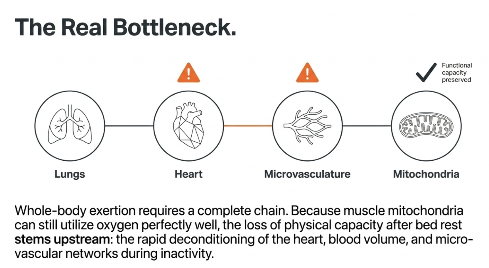
This study found the last link intact. The mitochondria could still use oxygen to make energy perfectly well. So if exercise capacity nonetheless falls after bed rest, the authors reason, the limiting factor is likely to lie earlier in the chain, in the delivery of oxygen rather than its use. And there is good reason to suspect exactly that, because the cardiovascular system is known to decondition quickly during bed rest. The heart's output drops, blood volume falls, and the microvascular and endothelial networks that regulate blood flow into working muscle lose some of their function. When the body lies still for days, the plumbing that delivers oxygen degrades faster than the engines that consume it.
This reframes post-illness weakness in a way that is both clarifying and, in a sense, encouraging. The conventional focus on mitochondrial decline implies that disuse damages the muscle's fundamental ability to produce energy, which sounds like a deep and hard-to-reverse problem. The alternative this study points to, that the main early loss is in cardiovascular and microvascular conditioning, points at a system that is famously responsive to being used. The cardiovascular deconditioning of bed rest is the same deconditioning that physical activity reverses, often relatively quickly. If the engines are fine and the problem is mostly the delivery system, then the path back runs through the kind of reconditioning the body is well equipped to do.
Two cautions keep this honest. First, this study did not measure the cardiovascular side. Its conclusion about where the functional loss comes from is an inference drawn from finding the mitochondria intact, a well-reasoned inference consistent with a large body of bed-rest research, but an inference rather than a direct measurement in these participants. Second, ten days is a short window. The authors are clear that longer inactivity may eventually overwhelm the compensations the mitochondria mounted here, and that the mitochondrial picture could look worse with more time in bed. What this study captures is the early phase, when the engines are still holding and the delivery system is likely the first thing to give.
What the Study Can and Cannot Tell Us
This is a carefully executed study with an unusually complete set of methods, but its scope is bounded, and reading it well means holding those bounds in view.
The sample was small: ten men. The authors are candid about this and defend it on practical grounds, bed rest studies are extraordinarily demanding to run, requiring participants to be housed and monitored in a hospital ward for the full duration, which is expensive and logistically heavy, and more so with older participants who need close medical oversight. They note that other well-regarded bed rest studies have used similar numbers, which is true. But a study of ten people still has limited statistical power, is more vulnerable to the influence of individual variation, and cannot speak to how consistently these findings would hold across a larger and more diverse group. Several analyses dropped to eight or nine participants after statistical outliers were removed, which tightens the findings but further shrinks already small numbers.
It studied only men. Whether the same mitochondrial response to disuse occurs in older women is unknown and genuinely important, because muscle metabolism and its age-related changes can differ between the sexes, and women are at least as affected by the frailty that follows periods of inactivity. The findings describe older men and should not be assumed to generalize to older women without direct study.
It captured a single snapshot in time. The muscle was examined once at the end of the ten days, which reveals where things stood at that point but not how they got there or where they were heading. The early days of the response, and what happens with longer disuse or during recovery, are invisible to this design. This matters especially for the study's more hopeful notes: the compensations the mitochondria mounted, the preserved efficiency, the tightly controlled exhaust under demand, are a tenth-day picture, and the authors are explicit that longer inactivity could overwhelm them.
Some of the key conclusions rest on gene expression rather than on function or protein. The antioxidant-defense story, the heart of the reframe, comes substantially from the transcriptomic data, the pattern of which genes were switched up or down. Gene-expression changes are a strong indicator of what the cell is programming itself to do, but they are not the same as a measured change in the actual antioxidant enzymes or in realized damage. The recycling story carries the same caveat, since the genes shifted while the key proteins did not. The authors present these as likely mechanisms supported by converging evidence, not as fully nailed-down causal chains, and the article follows them in that.
And the central claim about where functional loss comes from, the cardiovascular reframe, was not tested here at all. It is an inference from finding the mitochondria intact, well grounded in the broader bed rest literature but not measured in these participants. The study is strong evidence that the muscle mitochondria are not the early problem. It is suggestive, not conclusive, about what the early problem actually is.
None of this undercuts the core, well-supported finding: that ten days of disuse in older muscle raised oxidative stress and thinned the mitochondrial population while leaving energy production intact, placing the early damage in redox balance and mitochondrial mass rather than in the engines. But it is one small, acute, single-sex, single-timepoint study, and it is best read as a sharp and valuable piece of a larger picture rather than the final word on any part of it.
What This Means
For anyone thinking about how to age well, and especially for anyone caring for an older parent or heading into their own later decades, this study carries a few lessons that are practical even though its subject is a controlled laboratory bed rest.
The first is that short periods of inactivity are not harmless, and the damage is faster than most people assume. Ten days, the length of an ordinary hospital stay or a rough bout of illness, was enough to measurably raise oxidative stress and thin the mitochondrial population in older muscle. This happened in healthy older men under controlled conditions, which means it is likely worse, not better, in the frailer, sicker people who actually end up in hospital beds. The practical implication is one that rehabilitation medicine has been moving toward for years and that this study reinforces at the molecular level: avoiding unnecessary bed rest, and getting older patients moving as early and as safely as possible during and after illness, is not a nicety. It is protecting the muscle from real, measurable harm that accrues day by day.
The second lesson is about where the harm sits, and therefore what actually helps. The damage was in redox balance and in the number of mitochondria, not in the engines' capacity to produce energy, and the early functional loss likely comes from cardiovascular deconditioning rather than from the muscle itself. Both of those point in the same direction: toward physical activity as the lever. Movement is the most established way to support the body's antioxidant defenses, to stimulate the replacement of lost mitochondria, and to recondition the cardiovascular system that delivers oxygen to muscle. It is worth being explicit about what this does not point to, which is antioxidant supplements. It is tempting to read "oxidative stress rose" as "take antioxidants," but the research on that is discouraging, high-dose antioxidant supplements have generally failed to help and can even blunt the beneficial adaptations that exercise produces. The body's own antioxidant systems are best supported by using the muscle, not by flooding it with supplemental antioxidants. The lever is activity, not a pill.
The third lesson is the one that reaches beyond bed rest, and it is about the invisibility of all this. Everything the study measured, the rising oxidative stress, the thinning mitochondria, the shifting genetic program, was happening silently inside the muscle, detectable only because researchers took biopsies and ran them through a battery of molecular tests. The men did not feel their antioxidant genes downregulating. This is the uncomfortable truth about aging at the cellular level: the changes that matter most accumulate quietly, well before they surface as weakness or disease, which makes them easy to ignore until they are harder to reverse. The only way to know what is actually happening to your body, rather than guessing from how you feel, is to measure it.
That principle is the premise behind Healthspan's BioAge+, which measures the biological and metabolic markers of how a person is aging, turning the kind of changes that would otherwise stay invisible into something you can actually see and track over time. This study is a vivid reminder that the body's condition is written in its biology long before it is written in how a person looks or feels, and that the processes driving aging move whether or not anyone is watching. Knowing where you actually stand, and watching how it changes as you train, recover, and live, is the difference between managing your aging on evidence and managing it on assumption.
Conclusion: The Engine Was Never the Problem
The intuitive story about disuse and the aging body is that stillness breaks the muscle's engines, that the energy-producing machinery falters, and that this is why older people grow weak after a spell in bed. This study, with its unusually thorough look inside the muscle, tells a different and more precise story. After ten days of bed rest, the engines of these older muscles were not broken. They were intact, and by one measure each was running slightly better than before. Whatever was going wrong, it was not a failure to make energy.
What had gone wrong was subtler and, in its way, more instructive. The muscle's antioxidant defenses had been turned down, letting oxidative stress rise even though the mitochondria themselves were not producing more exhaust than their condition warranted. And the mitochondrial population had thinned, the fleet reduced, as the muscle cleared out old units faster than it built new ones. The early damage of disuse, in other words, was a problem of maintenance and balance, of the systems that keep the muscle clean and well stocked, rather than of the core capacity to produce power. And the weakness people actually feel after such a period likely comes from somewhere else entirely, from a cardiovascular system that deconditions faster than the muscle's engines do.
That reframing changes the response it calls for. If the problem were failing engines, the outlook would be grim, because the capacity to produce energy is fundamental and not easily rebuilt. But a redox imbalance, a thinned mitochondrial fleet, and a deconditioned cardiovascular system are all problems the body is well equipped to reverse. Indeed, what this study captured was an early, still-compensating response, the kind of deconditioning that the evidence suggests is largely reversible once a person starts moving again, through the one intervention that addresses all three problems at once: movement. The study makes a quiet but strong case for why keeping older bodies active, and getting them active again quickly after illness, protects something real and measurable, and why the common instinct to simply rest and recover can, past a point, do its own kind of harm.
The deeper lesson is about what ages, and how quietly it happens. Ten days of stillness reshaped the molecular life of these muscles in ways no one could see or feel, shifting thousands of genes, raising oxidative stress, thinning the mitochondria, all beneath the surface of a body that looked unchanged. Aging works this way more often than not: not as a sudden visible failure, but as a slow accumulation of silent changes that only become undeniable once they have gone far enough to matter. The muscle's engines may hold longer than we feared. What needs tending is everything around them, and the tending has to start before the weakness shows.
- Motanova, E., Zuccarelli, L., Lysenko, E., Amoretti, S., Pirazzini, M., Rossetto, O., Brocca, L., Bottinelli, R., Pellegrino, M.A., Baldassarre, G., Gissi, C., Lippe, G., Gasparini, M., Šimunic, B., Pišot, R., Grassi, B. and Narici, M.V. (2026), Preserved mitochondrial respiration in presence of oxidative stress and reduced mitochondrial mass after 10-day bed rest in older adults. J Physiol, 604: 7772-7798. https://doi.org/10.1113/JP291588
Related studies