How Urolithin A Improved a Model of Heart Failure: Not Just by Enhancing Mitophagy, but by an Unexpected Route Through the Gut Microbiome
Prefer to listen? Hit play for a conversational, audio‑style summary of this article’s key points.
Half of all heart failure is the kind with almost no treatments, and it is not a pumping problem. In heart failure with preserved ejection fraction, or HFpEF, the heart squeezes normally but has become too stiff to relax and fill between beats. It accounts for roughly half of heart failure cases, rises with age and metabolic disease, and until recently had essentially no therapies targeting its underlying biology. The stiffness is the visible endpoint of a failure that begins inside the heart's cells.
A new study tested urolithin A, a gut-derived compound, in a mouse model of HFpEF, and it improved the disease on every measure. Diastolic filling improved, and cardiac hypertrophy, fibrosis, and lung congestion all decreased, along with the genes that drive scarring and thickening. Notably, this happened without the mice losing weight or eating less, which means the compound was acting on the heart's biology rather than simply reducing the metabolic stress.
Urolithin A is a gut-microbiome metabolite best known for activating mitophagy, the clearance of damaged mitochondria. It is produced from compounds in pomegranates, walnuts, and berries, though many people's gut bacteria cannot make it efficiently, which is why it is sold as a supplement. It is one of the few longevity compounds with any human trial data, though that data is for muscle and immune endpoints, not the heart.
The first mechanism restored mitochondrial quality control inside the heart cell. HFpEF hearts showed low AMPK activity and high mTOR, the configuration that shuts down cellular cleanup. Urolithin A reversed that balance, reactivated mitophagy, and restored both mitochondrial energy production and physical structure, with damaged, swollen mitochondria giving way to intact ones. The authors are careful not to claim AMPK activation is the single trigger, since improved mitochondria and restored cleanup likely reinforce each other.
The second mechanism ran entirely outside the heart, through the gut. Urolithin A remodeled the gut microbiome, reducing bacteria that produce ceramides, a class of lipids that become toxic to heart cells when they accumulate. Bacterial ceramide-synthesis genes fell, and circulating ceramides in the blood dropped. This is a genuinely systemic picture: a gut-derived compound looping back to lower a cardiotoxic lipid, independent of its effects inside the heart cell.
The gut-ceramide mechanism is correlational, not proven cause. The microbiome, the ceramide genes, and the blood ceramides all moved in the right direction together, but the study did not prove the gut change caused the cardiac improvement. The experiments that would establish causation, such as germ-free animals or microbiome transplants, were not done. It is a strong and coherent hypothesis, not a demonstrated pathway.
Human heart cells showed the anti-fibrotic effect, which is what makes the finding worth taking seriously. In human iPSC-derived cardiomyocytes under HFpEF-like stress, urolithin A suppressed the shift toward a scar-producing state. The authors were transparent about an odd artifact in the data, a stress-induced liver-gene signature they interpreted cautiously rather than spun, which is the kind of restraint that makes the rest of the work more credible.
This is a preclinical rationale, not evidence anyone should take urolithin A for their heart. The study was in mice, in a model capturing only part of human HFpEF, in young animals, testing prevention during ongoing injury rather than reversal of established disease, at doses far above what people take. No human heart has been tested. It is a strong reason to run a human cardiac trial, and not a reason to self-treat.
The bigger idea is that HFpEF may be treatable by repairing cellular maintenance rather than targeting the pump. This aligns with the rapamycin pilot Healthspan covered, where relieving the same mTOR signaling from the other direction improved diastolic filling in older adults. Two different compounds converging on the same premise, that the aging, failing heart is partly a heart whose cellular housekeeping has broken down, is more persuasive than either result alone.
Introduction: The Heart Failure With Almost No Treatments
For decades, heart failure meant one thing: a weak heart. A heart damaged by a heart attack or worn down by disease, unable to pump with enough force to move blood through the body. That version of the disease is now reasonably treatable, with a set of drugs that have been refined over thirty years and genuinely extend lives.
But roughly half of all heart failure is not that. In the other half, the heart pumps normally. Squeeze it and it contracts with full force, and on an imaging scan it can look entirely healthy. The problem is the opposite of weakness: the heart has become too stiff to relax. Between beats, a healthy heart expands and fills with blood, loose and elastic. A stiff one cannot. It fights the filling, pressure backs up into the lungs, and the person experiences the breathlessness and exercise intolerance of heart failure despite a heart that pumps fine.
This is called heart failure with preserved ejection fraction, or HFpEF, and it is one of the largest unsolved problems in cardiology. It accounts for approximately half of all heart failure cases. It rises with age, obesity, and metabolic disease, which means it is becoming more common, not less. And until very recently, it had essentially no treatments that addressed its underlying biology. The SGLT2 inhibitors, which Healthspan has covered extensively, are the one recent bright spot, and even their benefit in HFpEF is real but modest. For a condition this common, the therapeutic cupboard is close to bare.
Part of why HFpEF has been so hard to treat is that it is not really a plumbing problem. The stiffness and the fibrosis are the visible endpoint of something happening much deeper, at the level of cellular energy metabolism. The heart is the most energy-demanding organ in the body, and a growing body of evidence points to failing mitochondrial quality control as a central driver of the disease. The heart muscle cells accumulate damaged mitochondria they can no longer clear, energy production falters, and the tissue responds by stiffening and scarring. This framing has already motivated one line of longevity research Healthspan has covered: a recent proof-of-concept pilot in older men found that low-dose rapamycin, which relieves the same mTOR overactivation that suppresses cellular cleanup, produced measurable improvements in diastolic filling over eight weeks. The compound in this new study comes at the identical problem from a different angle.
A new study published in Experimental & Molecular Medicine tested whether a single gut-derived molecule could intervene at that deeper level. The molecule is urolithin A, already familiar to longevity researchers as one of the most studied activators of mitophagy, the cellular process that clears damaged mitochondria. In a mouse model of HFpEF, urolithin A improved the disease on multiple fronts. What makes the study worth reading is not just that it worked, but how: it acted through two largely independent mechanisms at once, one inside the heart cells and one running all the way out through the gut microbiome. It is one of the clearer demonstrations to date of how this compound operates across several biological levels simultaneously.
Before going further, the essential caveat. This is a mouse study, in a specific model, with the compound given during disease development rather than after the disease was established. It builds a mechanistic rationale. It does not show that urolithin A treats heart failure in people, and nothing here should be read as a reason to take it for that purpose. With that framing in place, the biology is genuinely interesting.
What HFpEF Actually Is
To follow what this study did, it helps to understand the disease it targeted, because HFpEF is one of the more counterintuitive conditions in medicine. The name itself contains the puzzle.
Ejection fraction is the fraction of blood in the left ventricle that gets pumped out with each beat. A healthy heart ejects a little more than half of what it holds, and ejection fraction is the standard number cardiologists use to measure whether a heart is pumping well. In the classic form of heart failure, this number drops. The muscle is weak, it cannot contract forcefully, and less blood gets pushed out with each beat. That is heart failure with reduced ejection fraction, and it is the version most people picture.
HFpEF is the paradox: the ejection fraction is preserved, meaning normal. Measure it and the heart appears to be pumping fine. And yet the person has unmistakable heart failure, with fluid in the lungs, breathlessness on exertion, swelling, and fatigue. The pumping was never the problem. The filling is.
A heartbeat has two phases. Systole is the contraction, when the ventricle squeezes and ejects blood. Diastole is the relaxation, when the ventricle releases, expands, and fills with the next volume of blood to pump. These are equally essential, but they place opposite demands on the muscle. Contraction requires the heart to generate force. Relaxation requires it to yield, to become momentarily loose and compliant so that blood can flow in easily and fill the chamber.
In HFpEF, the heart has lost the ability to yield. The muscle has become stiff and thick, and the walls no longer relax fully between beats. Picture the difference between a fresh rubber band and one that has hardened with age. The old band can still snap back when stretched, but it resists being stretched in the first place. A stiff ventricle behaves the same way. It can still contract and eject, but it fights being filled. To force enough blood into a chamber that resists expansion, pressure has to rise, and that elevated pressure backs up into the lungs, producing the congestion and breathlessness that bring people to the clinic.
What drives the stiffening is the convergence of several age-related and metabolic insults. Chronic high blood pressure forces the heart to work against resistance, and it responds the way any muscle does under sustained load, by thickening. Obesity, diabetes, and metabolic stress add a smoldering systemic inflammation that promotes fibrosis, the deposition of stiff collagen scaffolding between the muscle cells. The heart muscle cells themselves enlarge. The net result is a ventricle that is thicker, stiffer, more fibrotic, and progressively less able to perform the quiet half of its job.
This is why HFpEF has resisted the drugs that work for the other kind of heart failure. Those drugs largely help a weak heart pump or reduce the load it works against. None of them address stiffness, fibrosis, and impaired relaxation, which are problems of tissue quality rather than pumping force. To improve HFpEF at its root, an intervention has to reach the cellular processes that make the tissue stiffen in the first place. And that is where mitochondria enter the story.
Why Mitochondria Sit at the Center
The link between a stiffening heart and its mitochondria is not obvious, but it is central to why a mitophagy activator was worth testing at all.
Start with the energy problem. The heart never rests. It beats roughly once a second for a lifetime, and every one of those beats, the contraction and the relaxation both, costs energy. Relaxation is not passive. Actively pumping calcium back out of the machinery so the muscle can release its grip is one of the most energy-expensive things a heart cell does. All of that energy comes from mitochondria, the organelles that convert fuel into ATP, the cell's usable energy currency. The heart contains more mitochondria per cell than almost any other tissue, because its energy demand is relentless.
When mitochondria work well, the heart has the energy to both contract and relax cleanly. When they falter, the relaxation phase suffers first, because it is the more energetically demanding and less forgiving of an energy shortfall. This is the first thread connecting mitochondrial health to diastolic function: a heart cell short on energy is a heart cell that cannot fully relax.
The second thread is damage. Mitochondria are not just energy factories; they are also a source of cellular stress when they break down. A damaged mitochondrion produces less ATP and leaks reactive oxygen species, the corrosive byproducts of energy metabolism, which damage surrounding proteins, membranes, and DNA. A cell full of failing mitochondria is both underpowered and under chemical assault.
This is why cells have a dedicated quality-control system for mitochondria, and it is the process at the heart of this study: mitophagy. The word combines mitochondria with autophagy, the cell's general system for digesting and recycling its own damaged components. Mitophagy is the specialized version aimed at mitochondria specifically. When a mitochondrion is damaged beyond repair, it gets tagged for destruction, enveloped in a membrane, delivered to the cell's recycling compartment, and broken down into parts that can be reused. It is the cellular equivalent of pulling a broken machine off the factory floor before it causes an accident, and salvaging it for scrap.
A healthy heart cell runs this process continuously, clearing damaged mitochondria and maintaining a population of functional ones. The system is governed by a molecular decision point that recurs throughout this article. Two proteins sit at the center of it. AMPK is an energy sensor that switches on when the cell detects energy scarcity or stress, and one of its jobs is to activate autophagy, telling the cell to start recycling. mTOR is its counterpart, a growth regulator that switches on when nutrients are abundant, and it suppresses autophagy, telling the cell to build rather than recycle. The balance between these two, AMPK promoting cleanup and mTOR suppressing it, sets how actively a cell clears its damaged components.
In HFpEF, this balance tips the wrong way. Mitophagy becomes blunted. Damaged mitochondria that should be cleared instead accumulate, energy production drops, reactive oxygen species rise, and the cell slides into the dysfunction that drives hypertrophy and fibrosis. Recent work has established impaired mitophagy as a genuine contributor to HFpEF rather than a bystander, which is what makes the therapeutic logic tractable: if blunted mitophagy is part of what drives the disease, then a compound that restores mitophagy might interrupt it.
That is the precise hypothesis this study set out to test, and the compound it tested is one longevity research already knew well.
Urolithin A and the Logic of Testing It Here
Urolithin A is not a drug in the conventional sense. It is a compound your gut bacteria make, provided you have the right bacteria and the right raw material.
The raw material comes from ellagitannins, a class of polyphenols found in pomegranates, walnuts, raspberries, and a handful of other foods. You cannot absorb ellagitannins directly in any meaningful quantity. Instead, certain gut bacteria break them down through a series of steps, and urolithin A is the end product of that microbial processing. This arrangement has a consequence that turns out to matter for the whole field: not everyone's gut microbiome can perform the conversion. Estimates vary, but a substantial fraction of people, possibly the majority, produce little or no urolithin A even when they eat the foods that supply the precursor. This variability is part of why the compound is now sold as a direct supplement, bypassing the gut conversion entirely and delivering urolithin A itself.
What earned urolithin A its place in longevity research is a single, reproducible property: it activates mitophagy. This was established in a landmark 2016 study showing that urolithin A induced mitophagy and extended lifespan in the worm C. elegans, and improved muscle function in rodents. The finding has since been extended across species, and a randomized placebo-controlled trial in humans reported improvements in muscle endurance and markers of mitochondrial health. As mitophagy activators go, it is among the best characterized and one of the very few with any human trial data behind it at all.
Healthspan has covered urolithin A before, including its mechanistic relationship with rapamycin, and the two compounds make an instructive pair. Rapamycin activates mitophagy from the top down, by inhibiting mTOR and lifting its suppression of autophagy. Urolithin A works from a different direction, promoting mitophagy more directly and, as this study shows, engaging AMPK. Same destination, different roads. That distinction becomes relevant later, because it is part of why the two might eventually be complementary rather than redundant.
The logic of this study now assembles cleanly. HFpEF appears to be driven in part by failing mitochondrial quality control. Urolithin A is a well-characterized activator of mitochondrial quality control. Nobody had yet tested whether it could help in HFpEF specifically, a disease defined by metabolic stress and structural remodeling rather than the muscle-wasting contexts where urolithin A had mostly been studied. The question was direct: give urolithin A to an animal with HFpEF, and does the heart get better?
How the Study Was Built
Testing that question requires a version of HFpEF you can study in the laboratory, and this is the first place the study's design deserves scrutiny, because modeling HFpEF in a mouse is genuinely difficult.
Most heart failure models produce the weak-heart kind, by damaging the muscle directly. HFpEF is harder to reproduce, because its defining feature is preserved pumping with impaired relaxation, and that arises from the convergence of metabolic and vascular stress rather than from a single injury. The field's current best approach is what researchers call a two-hit model, and it is what this study used.
The two hits were a high-fat diet and a compound called L-NAME. The high-fat diet supplies the metabolic stress, producing obesity, disordered metabolism, and the systemic conditions that predispose to HFpEF. L-NAME supplies the vascular stress: it blocks the production of nitric oxide, the molecule blood vessels use to relax and dilate, which drives up blood pressure and mimics the vascular dysfunction that accompanies the human disease. Together, over eight weeks, the two hits produce mice with the recognizable features of HFpEF: preserved ejection fraction, impaired diastolic relaxation, cardiac hypertrophy, and fibrosis. The researchers confirmed the phenotype at the eight-week mark before proceeding, checking that ejection fraction remained normal while a measure of diastolic filling called the E/A ratio had become abnormal.
The design choice that matters most for interpretation comes next. After confirming the HFpEF phenotype at week eight, the researchers continued the high-fat diet and L-NAME for another twelve weeks, and during that period they added urolithin A to the diet for the treatment group, at one of two doses. In other words, the disease-causing insults never stopped. Urolithin A was given alongside ongoing injury, not after the injury had ended.
This makes the study a model of intervention during continued disease progression. It is a fair test of whether urolithin A can blunt or hold back the disease while the causes are still active, which is arguably the realistic clinical scenario, since a patient's hypertension and metabolic stress do not vanish when treatment begins. But it is not a test of whether urolithin A can reverse established, stable disease, and it is worth being precise about that distinction throughout. The honest framing is that urolithin A improved the trajectory of an actively developing disease, not that it reversed a settled one.
One other feature of the design is worth noting in its favor. The researchers did not rely on the mouse alone. They paired the animal work with experiments in human cardiomyocytes derived from induced pluripotent stem cells, cells reprogrammed from human donors and coaxed into becoming heart muscle. This lets them ask whether the mechanisms seen in the mouse also operate in human cells, which is a meaningful step up from a mouse-only study, even though these lab-grown cells are immature and not equivalent to an adult human heart.
With the model established, the results fall into two threads: what urolithin A did to the heart, and how it did it.
What Urolithin A Did to the Heart
The top-line result is that urolithin A improved HFpEF across every major dimension the researchers measured, from whole-organ function down to the molecular signature of the disease.
The most important measure is diastolic function, since that is what defines HFpEF. Using echocardiography, the researchers tracked how well the ventricle filled between beats. The E/A ratio, an index of filling that had become abnormal as the disease developed, moved significantly back toward normal in the treated mice. Because diastolic function is the thing HFpEF breaks, this is the result the rest of the study has to explain.
The structural findings tracked with the functional ones. Untreated HFpEF hearts showed the expected remodeling: thickened ventricular walls, increased heart mass, and the fluid backup into the lungs that reflects elevated filling pressure. Urolithin A, delivered in the diet at 250 or 500 mg per kilogram per day across the twelve-week treatment window, attenuated all of it. Wall thickness and heart mass came down, and the pulmonary congestion, measured by lung weight, normalized, which is a physiologically meaningful sign that filling pressures had eased.
Fibrosis showed the same pattern. Fibrosis is the deposition of stiff collagen scaffolding between heart muscle cells, and it is one of the primary physical reasons an HFpEF heart cannot relax. Staining the heart tissue revealed extensive collagen deposition in untreated animals, both between muscle cells and around blood vessels, and urolithin A markedly reduced it. At the genetic level, the drivers of fibrosis and hypertrophy told a consistent story: genes including Tgfb1, Col1a1, and Ctgf, which orchestrate collagen deposition, were elevated in HFpEF and brought back down by urolithin A, as were the hypertrophy genes Myh7 and Nppb.
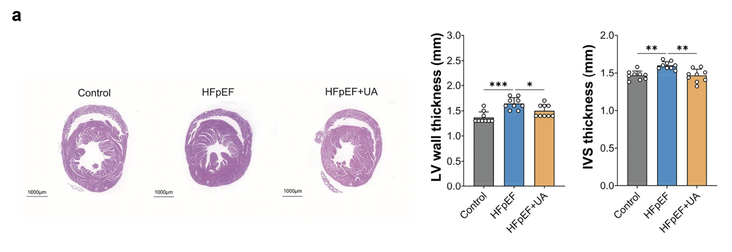
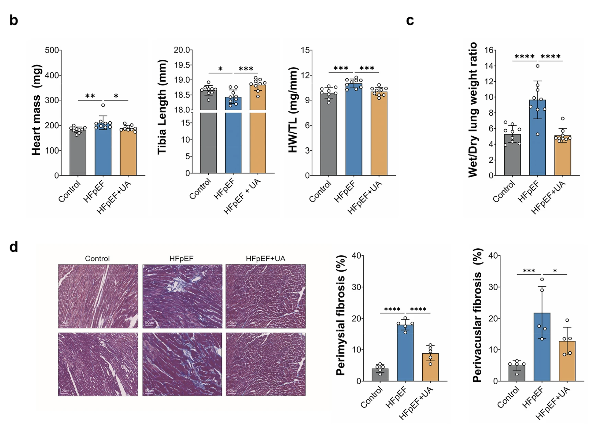
Figure 1: Urolithin A reduces fibrosis and hypertrophy. Tissue staining shows collagen deposition between muscle cells and around vessels, extensive in untreated HFpEF hearts and substantially reduced with urolithin A. The fibrosis genes Tgfb1, Col1a1, and Ctgf and the hypertrophy genes Myh7 and Nppb, all elevated in HFpEF, were brought back down by treatment.
One negative result is as important as any of the positive ones. Urolithin A produced none of these benefits by making the mice thinner or changing what they ate. Body weight, food intake, and body composition, including both lean and fat mass, were unchanged between the treated and untreated HFpEF groups. This matters because the easy explanation for any improvement in a high-fat-diet model is that the intervention simply reduced the metabolic insult, made the animals leaner, and improved the heart secondarily. That explanation is ruled out here. The heart improved while the metabolic stressor stayed constant, which means urolithin A was acting on the heart and its supporting systems rather than on the mouse's weight.
So the effect is real and it is not an artifact of weight loss. The substance of the study is the mechanism, and this is where its two independent threads separate. One runs inside the heart cell. The other runs through the gut.
Mechanism One: Restoring Mitophagy Inside the Heart Cell
The first mechanism is the one the study was designed around, and it plays out exactly along the AMPK and mTOR decision point described earlier.
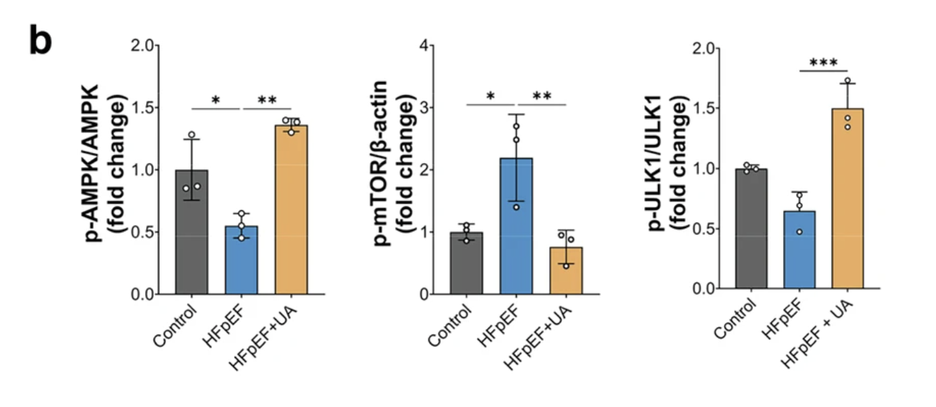
The researchers began by looking at that signaling balance in the heart tissue. In untreated HFpEF hearts, it was tipped the wrong way: AMPK activity, the signal that promotes cellular cleanup, was reduced, while mTOR activity, the signal that suppresses it, was elevated. This is the molecular configuration of a cell that has stopped recycling and cannot easily restart. Urolithin A reversed it, raising AMPK activity and lowering mTOR, and restoring activity in ULK1, the enzyme that sits just downstream of AMPK and actually initiates the construction of the machinery that engulfs damaged components.
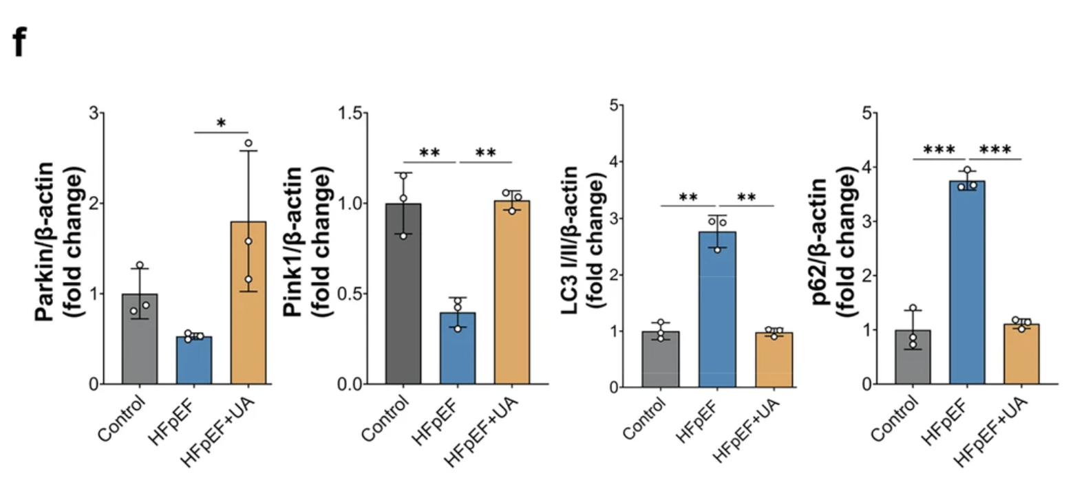
With the signaling restored, the question was whether mitophagy itself resumed. The researchers approached this from several angles, which matters, because mitophagy is notoriously easy to misjudge from any single measurement. They examined the canonical mitophagy proteins, PINK1 and Parkin, which tag damaged mitochondria for removal, and found both restored by urolithin A treatment. They tracked markers of autophagic flux, the throughput of the recycling system read through the proteins LC3 and p62, and saw the accumulation that signals a jammed system give way to normal turnover.
The most direct evidence came from a reporter called mt-Keima, a fluorescent tool that changes color depending on whether a mitochondrion is sitting in the neutral environment of the cell or has been delivered into the acidic environment of the recycling compartment. It provides a visual readout of mitophagy actually happening rather than an inference from protein levels. Under HFpEF-like stress, the signal indicated stalled mitophagy. Urolithin A shifted it toward active mitochondrial clearance, comparable to the effect of a chemical compound used as a positive control specifically because it forces mitophagy.
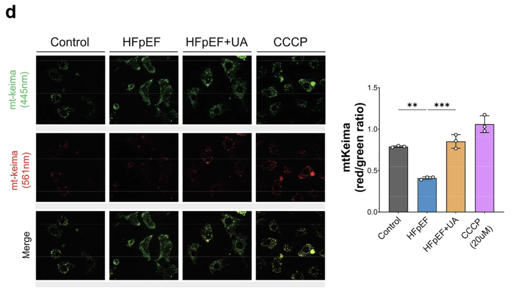
Two functional confirmations close the loop. First, respiration: using a technique that measures how much oxygen cells consume to make energy, the researchers found that HFpEF had suppressed basal respiration, ATP-linked respiration, and maximal respiratory capacity, and that urolithin A restored all three, along with the cell's spare respiratory capacity, its reserve for meeting surges in demand. The heart cells were making energy properly again. Second, structure: under electron microscopy, which shows mitochondria directly, untreated HFpEF hearts contained the visible signature of the disease, swollen and fragmented mitochondria with disrupted internal architecture. Treated hearts showed mitochondria with preserved structure and intact cristae, the folded internal membranes where energy production happens.
Alongside clearance, the balance of mitochondrial fusion and fission had also gone wrong in HFpEF, and urolithin A corrected it. Mitochondria continually merge and divide, and the proteins governing that balance were skewed in the diseased hearts: MFN1, which drives fusion, was reduced, while DRP1, which drives fission, was elevated, a configuration that fragments the mitochondrial network. Urolithin A restored both toward normal. This matters because fragmentation is itself a marker of mitochondrial distress, so correcting it reflects a mitochondrial population returning to health rather than one being cleared and rebuilt under duress.
So the first mechanism is complete and internally consistent, from signaling to flux to function to structure: urolithin A reactivated the AMPK-driven mitophagy that HFpEF had blunted, the cell resumed clearing its damaged mitochondria, and mitochondrial function and structure recovered.
The study is careful at exactly the point where it would be easy to overreach, and the article should be too. It is tempting to draw a clean arrow: urolithin A activates AMPK, AMPK drives mitophagy, mitophagy fixes the heart. The authors explicitly decline to claim that the AMPK activation is the single upstream trigger. AMPK responds to the cell's energy state, so improved mitochondrial function could be a cause of the AMPK activation as easily as a consequence of it. The likeliest reading is a feedback loop rather than a one-way street, where restored energy and restored cleanup reinforce each other, and the authors note that establishing strict causation would require directly blocking AMPK to see whether the benefit disappears. This is the kind of restraint that makes the rest of the paper more credible, not less.
Mechanism Two: The Gut-Ceramide Axis
The second mechanism is the one that makes this study genuinely novel, because it runs outside the heart entirely, beginning in the gut and traveling to the heart through the bloodstream.
The starting observation is that HFpEF is increasingly understood as a systemic metabolic disease, not a purely cardiac one, and part of that systemic picture involves the gut microbiome. The community of bacteria in the intestine produces a vast array of molecules that enter circulation and affect distant organs, and disturbances in that community have been linked to heart failure. The researchers asked whether urolithin A, itself a product of gut bacteria, also acts back on the gut in a way that matters for the heart.
To find out, they sequenced the gut bacteria of the mice and measured lipids in their blood. The HFpEF mice showed the disordered microbiome that accompanies metabolic disease: reduced diversity and a shift in composition, including an expansion of several bacterial groups associated with producing ceramides. Urolithin A restored microbial diversity and reduced those ceramide-associated bacteria toward normal levels.
Ceramides are the link that makes this cardiac. They are a class of lipids that do far more than store energy or build membranes. When they accumulate in excess, they become actively toxic to cells, a phenomenon called lipotoxicity. Ceramides impair mitochondrial function, activate inflammatory machinery, and push cells toward the fibrotic, stressed states that characterize a failing heart. Elevated ceramides are an established marker of cardiovascular risk, and there is evidence they are not merely a marker but a driver. So a molecule that lowers the body's ceramide burden is plausibly doing something protective for the heart.
The study traced the connection across three levels. First, the microbiome: urolithin A reduced the bacterial groups whose genes encode ceramide-building enzymes. Second, the bacterial gene function: metagenomic analysis confirmed that the genes for ceramide synthesis, elevated in HFpEF, were suppressed by urolithin A. Third, the blood: plasma lipid measurement confirmed that circulating ceramides, elevated in the HFpEF mice, fell with treatment. The chain runs from a shift in which bacteria are present, to a shift in what those bacteria are genetically equipped to make, to a measurable drop in the toxic lipid in the blood.
That is a coherent and, in its details, well-supported story: urolithin A remodels the gut microbiome, the remodeled microbiome produces fewer ceramides, circulating ceramides fall, and the heart is exposed to less lipotoxic stress. Crucially, this pathway is independent of the mitophagy mechanism. One works inside the heart cell restoring quality control; the other works through the gut lowering a systemic toxin. Two different routes, converging on the same diseased organ.
But this is also the mechanism where the honest caveat has to be loudest, and the authors are appropriately clear about it. Everything in this chain is correlational. The microbiome changed, the ceramide genes changed, and the blood ceramides fell, all in the same direction at the same time, but the study did not prove that the microbiome change caused the ceramide drop, or that the ceramide drop caused the cardiac improvement. The associations are strong and biologically sensible, but association is not causation. Establishing that the gut is genuinely driving the cardiac benefit, rather than changing in parallel with it, would require experiments the study did not perform: raising the mice germ-free, depleting their microbiomes, or transplanting the treated microbiome into untreated animals to see whether the protection transfers with it. Until then, the gut-ceramide axis is a compelling and coherent hypothesis rather than a demonstrated causal pathway.
That distinction matters for how much weight the mechanism can bear, but it does not diminish what makes it interesting. Even as a correlation, the finding that a gut-derived compound loops back to reshape the gut and lower a cardiotoxic lipid is a genuinely systemic picture of how a single molecule might act on a disease this diffuse.
The Human-Cell Check
Both mechanisms so far come from mice, and the gap between a mouse heart and a human one is exactly where most promising cardiac findings fail. To probe whether any of this might carry over, the researchers turned to human heart cells, and this is where the study earns some of its credibility.
They used cardiomyocytes derived from human induced pluripotent stem cells. The starting material is ordinary human cells, often skin or blood, reprogrammed back into a stem-like state and then directed to become beating heart muscle cells. The result is human cardiac tissue in a dish, carrying a human genome and human cellular machinery, without anyone having to sample a living heart. The researchers confirmed the cells were genuine cardiomyocytes by their expression of the proteins that define heart muscle, then exposed them to the same kind of metabolic and nitrosative stress used to model HFpEF in the mice.
Under that stress, the human cells drifted toward disease. Using single-nucleus RNA sequencing, a technique that reads gene activity one cell at a time, the researchers watched the population shift toward an inflammatory, fibrotic, hypertrophic expression profile, the cellular fingerprint of the same remodeling seen in the whole animal. Urolithin A blunted the shift. The inflammatory and profibrotic programs the stress switched on were substantially quieted by treatment.
The single-cell resolution let them see something a bulk measurement would have missed. As the cells were stressed, a distinct subpopulation emerged that was drifting toward a heavily fibrotic state, which the researchers labeled a fibrogenic cluster. Urolithin A did not just prevent this population from expanding; it reduced it below the level seen in unstressed control cells, suggesting the compound actively suppresses the fibrogenic transition rather than merely holding it in check. Tracing the cells along their trajectory of change confirmed the pattern: the march toward a scar-producing state was interrupted by treatment.
There is one wrinkle in this data that the authors handle with commendable transparency, and it is worth relaying because it illustrates the caution the whole study is written with. Under stress, a small population of the heart cells began expressing genes normally associated with liver cells, including albumin. Taken at face value, that is biologically implausible; heart cells do not become liver cells. Rather than ignore the anomaly or spin it, the authors explain it directly. Lab-grown iPSC cardiomyocytes are immature and prone to unstable gene expression, stress can push cells into aberrant transcriptional states, and single-nucleus sequencing is susceptible to technical artifacts like stray RNA from other cells. They interpret the liver-like signature as a stress-induced aberration rather than a real cell type, which is almost certainly correct, and they flag it rather than bury it. That the treated cells showed less of this aberrant state than the stressed ones fits the broader pattern, but the authors do not lean on it, and neither should a reader.
What the human-cell work establishes is modest but real. It does not show that urolithin A helps human patients, and iPSC cardiomyocytes are a long way from an aging human heart. But it demonstrates that the anti-fibrotic effect is not a quirk of mouse biology, that human heart cells under HFpEF-like stress respond to urolithin A in the same direction the mouse hearts did. That is the kind of cross-species consistency that makes a preclinical finding worth taking seriously, without pretending it is more than it is.
Limitations of the Study We Need to Consider
The distance between this study and a bottle of urolithin A on a shelf is considerable, and the limitations are worth stating plainly, because this is precisely the kind of result that gets over-read.
It is a mouse study. Everything in the animal work happened in mice, whose cardiac physiology, metabolic rate, and lifespan differ from ours in ways that matter. The history of cardiology is full of interventions that improved mouse hearts and did nothing for human ones. The human-cell work strengthens the case but does not close this gap, because lab-grown cardiomyocytes are immature and isolated from the whole-body context of a real heart.
The disease model is not the human disease. The two-hit model reproduces key features of HFpEF, but human HFpEF is not one disease. It is a heterogeneous syndrome arising from many combinations of hypertension, obesity, diabetes, kidney disease, and aging, and a model that captures some of its features in a young mouse over a few months cannot represent that full complexity. Notably, the mice were young. Human HFpEF is largely a disease of older people, and aging itself changes how the heart and the microbiome respond to intervention.
It was prevention during ongoing injury, not reversal of established disease. This is the distinction that most changes how the result should be read. Urolithin A was given while the high-fat diet and the nitric oxide blocker continued, so it was tested against a disease still actively developing, not one already established and stable. The study shows urolithin A can blunt the progression of HFpEF while the causes are still operating. It does not show that urolithin A can reverse fibrosis and stiffness that have already set in, which is the situation an actual patient is usually in by the time they are diagnosed.
The doses were high, and do not translate simply. The mice received urolithin A at 250 to 500 mg per kilogram of body weight per day. Human urolithin A trials typically use a fixed dose on the order of 500 mg total per day, not per kilogram. Dose does not scale linearly across species, and mice metabolize compounds faster than humans, so the mouse doses cannot be read directly as a human target. But the gap is a reminder that the exposures producing these effects in mice are not obviously matched by the amounts people actually take.
The microbiome findings are correlational. As detailed above, the gut-ceramide mechanism rests on associations that moved in the right direction together, not on experiments proving the gut drove the cardiac benefit. The causal tests, germ-free animals, microbiome depletion, or fecal transplant, were not done.
Key metabolic variables were not measured. The study did not assess glucose tolerance or insulin sensitivity, so the extent to which systemic metabolic changes contributed to the cardiac effects cannot be determined. In a high-fat-diet model, that is a meaningful gap.
Durability is unknown. The study does not address whether the benefits persist after urolithin A is stopped, or whether continuous use would be required. For anything that would be taken as a long-term supplement or therapy, that is a central open question.
None of these limitations make the study less interesting as mechanism. They make it clear that the study is what its authors say it is, a preclinical rationale for investigating urolithin A in HFpEF, and not evidence that anyone with the condition should take it.
What This Means
Three things are worth taking from this study, at three different distances from the clinic.
The mechanism is the real contribution. What distinguishes this work is not that urolithin A helped an HFpEF mouse, but that it did so through two independent routes at once, one restoring mitochondrial quality control inside the heart cell, the other lowering a cardiotoxic lipid through the gut. Most interventions are studied as single mechanisms. Seeing one molecule act on a diffuse, multi-system disease from two directions simultaneously is a more realistic picture of how an intervention might work against something as heterogeneous as HFpEF, where no single lever is likely to be enough. Whether both routes prove causally necessary is unresolved, but the framing itself, one compound reaching a disease through parallel systems, is the useful idea.
It strengthens the metabolic model of HFpEF. The finding reinforces a shift in how the disease is understood, away from a purely mechanical problem of a stiff heart and toward a problem of cellular energy management. If restoring mitochondrial quality control improves the disease, that is evidence mitochondrial failure was part of what drove it. This aligns with the rapamycin pilot Healthspan covered earlier, where relieving mTOR overactivation, the same signaling node urolithin A engages from the other side, produced measurable improvements in diastolic filling in older adults. Two different compounds, converging on the same underlying idea, that the aging, failing heart is in part a heart whose cellular maintenance has broken down. That convergence is more persuasive than either result alone.
The honest clinical position is restraint. Urolithin A is genuinely interesting for cardiometabolic aging, and it is unusual among longevity compounds in having human trial data, though that data is for muscle and immune endpoints, not the heart. For HFpEF specifically, the evidence is one preclinical study, in mice, testing prevention rather than reversal, at doses that do not map cleanly onto human use. That is a reason for researchers to design a human cardiac trial. It is not a reason for anyone with HFpEF, or worried about it, to start taking urolithin A expecting cardiac benefit. The gap between a mechanistic result in mice and a validated therapy in people is exactly where most candidates fail, and the appropriate stance is interest, not action.
The larger point connects this study to a theme that runs through much of Healthspan's coverage. The most interesting longevity interventions increasingly work not by targeting a single disease but by restoring a fundamental cellular process, mitochondrial quality control, autophagy, metabolic signaling, that degrades across many tissues with age. Urolithin A in HFpEF is one instance of that broader pattern: treat the upstream cellular failure, and a downstream age-related disease may improve as a consequence. It is a promising idea with real mechanistic support and, as yet, no proof in human hearts.
Conclusion: Treating the Cell, Not Just the Symptom
Heart failure with preserved ejection fraction looks, on the surface, like a mechanical problem. The heart is too stiff to fill, so the intuitive fix would be something that makes it more supple. But the stiffness is downstream. It is the visible endpoint of heart muscle cells that can no longer manage their own energy, clear their own damaged components, or resist the metabolic and inflammatory stress pressing on them from the rest of the body. By the time the ventricle has stiffened, the failure has already happened at the level of the cell.
This study is interesting because it intervenes at that deeper level, and because it does so from two directions at once. Inside the heart cell, urolithin A restored the mitophagy that clears damaged mitochondria, reactivating a quality-control system that HFpEF had shut down. Outside the heart, it remodeled the gut microbiome and lowered the circulating ceramides that poison cardiac tissue from the bloodstream. One molecule, two independent routes, converging on the same failing organ. That the disease improved across every dimension the researchers measured, function, structure, fibrosis, and molecular signature, without the mice losing any weight, indicates the compound was acting on the biology of the disease rather than on its metabolic backdrop.
The caveats are not incidental, and they set the ceiling on what can be claimed. This was a mouse study using a model that captures only part of human HFpEF, in young animals rather than old ones, testing whether the compound could hold back a disease still actively forming rather than reverse one already established, at doses well above what people take. The most novel mechanism, the gut-ceramide axis, rests on correlation rather than proven cause. And no human heart has been tested. What the study delivers is a mechanistic rationale, coherent and multi-layered and reinforced in human cells for its anti-fibrotic effect, for asking whether urolithin A could help people with HFpEF. It does not deliver an answer, and it is not a reason to self-treat.
What makes the result worth attention anyway is the shape of the idea behind it. HFpEF has resisted treatment because the drugs built for the other kind of heart failure address pumping, and HFpEF is not a pumping problem. An intervention that instead restores the cell's own maintenance machinery is aimed where the disease actually originates. Whether urolithin A specifically becomes part of the answer will be settled in human trials that have not yet been run. But the strategy it points toward, treating age-related disease by repairing the cellular processes that fail underneath it, is one of the more promising directions in longevity medicine, and this study is a careful, honest example of what that strategy looks like when it is tested rather than assumed.
Related studies